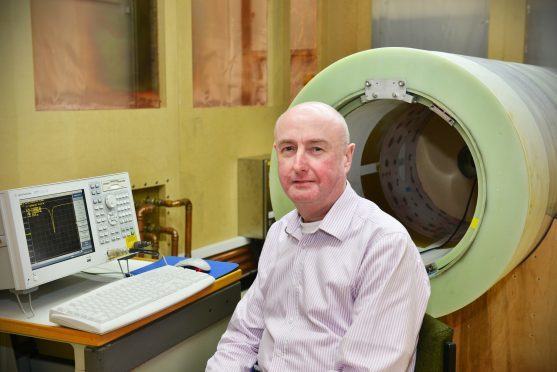
Experts at Aberdeen University are leading the way in developing state-of-the art MRI scanners to identify major diseases such as cancer and osteoarthritis more quickly.
The development of the specialist scanners has moved a step closer, following the award of a 6.6million grant from the European Commission.
Aberdeen University built and used the first clinical MRI machine to scan a man 35 years ago.
Now, the new method, which is thought to be the first of its type in the world, will reveal far more detailed information about diseases including osteoarthritis, cancer and dementia.
Current MRI machines use just one magnetic field, but fast field cycling MRI scanners will allow patients to be passed through thousands of possible variations whilst they are inside the machine, producing more information about the inside of their body.
Professor David Lurie of Aberdeen University, said: “There are a whole range of diseases where FFC-MRI could benefit diagnosis and monitor the success of treatment.
“Early tests show it can measure changes in cartilage in osteoarthritis and our research has also shown that there are changes visible in cancer.
“We believe there may be changes in many other conditions too, such as in the brain which are associated with dementia.
“The signals measured by FFC-MRI are a function of what’s happening on a molecular level in tissues which is where disease begins, but we’re not certain what is happening, and what’s causing these changes.
“That is what this new project will aim to explore, as well as developing techniques to improve the scanners and to provide better quality images of the body.”
Aberdeen University will oversee the four-year project which involves nine teams spanning six countries.
The Aberdeen-led consortium of groups from universities, research organisations and industrial partners fought off stiff competition for the funds from across Europe.
The findings from the new project will be brought together in four years’ time and passed on to the medical imaging industry.
