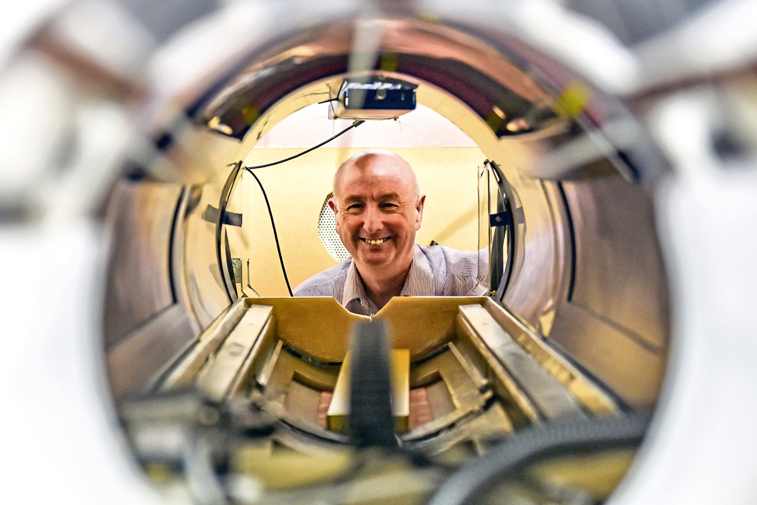
One of the most revolutionary pieces of medical equipment was pioneered under NHS Grampian almost 40 years ago.
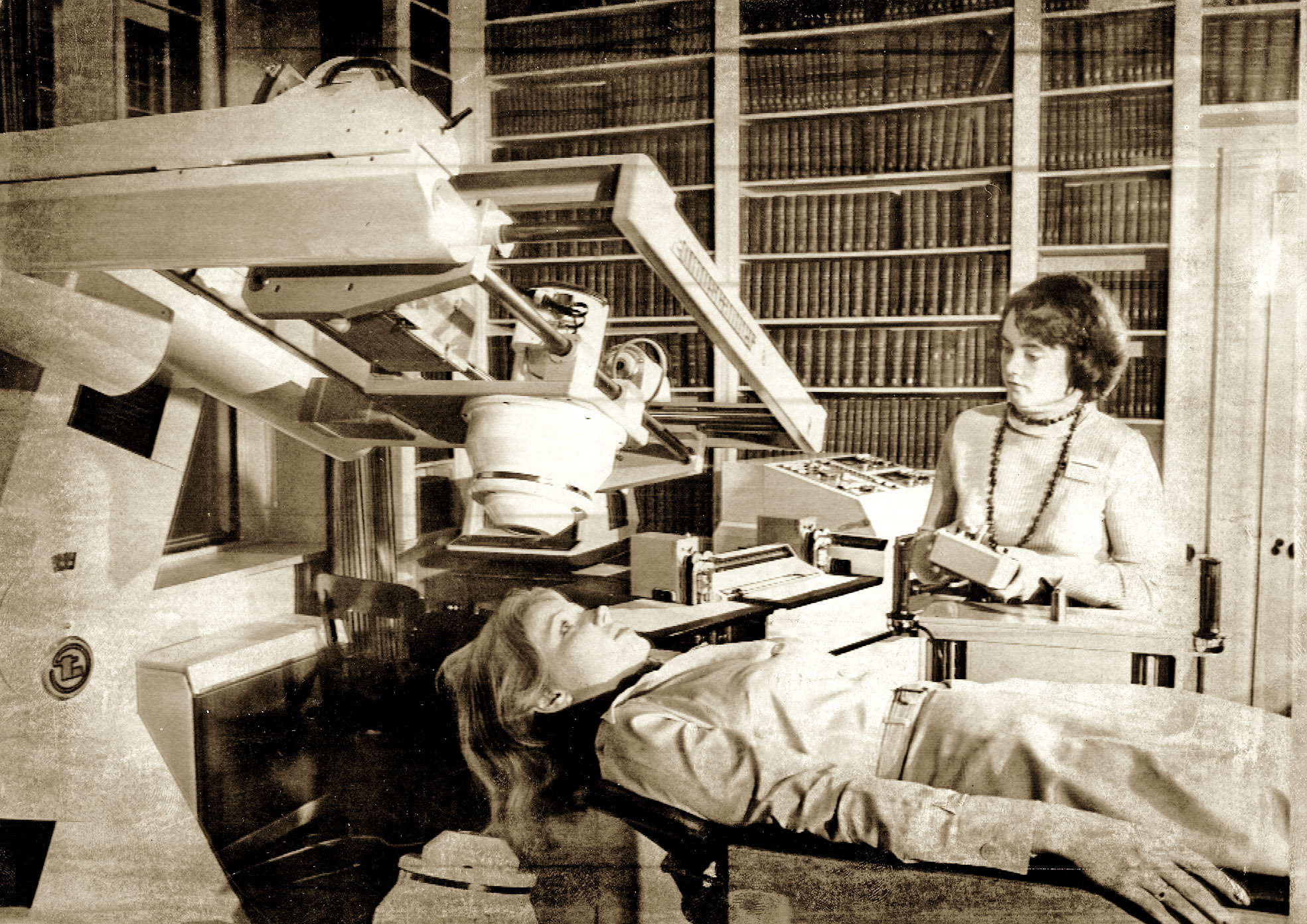
On August 28 1981, the clinical use of Magnetic Resonance Imaging (MRI) was a world first at Aberdeen Royal Infirmary.
Its invention, also known as the Mark-1, was revolutionary because, unlike X-rays, it has no risk of radiation.
An elderly Fraserburgh man became the first patient in the world to have a whole body scan.
It picked up tumours in his liver which would eventually claim his life.
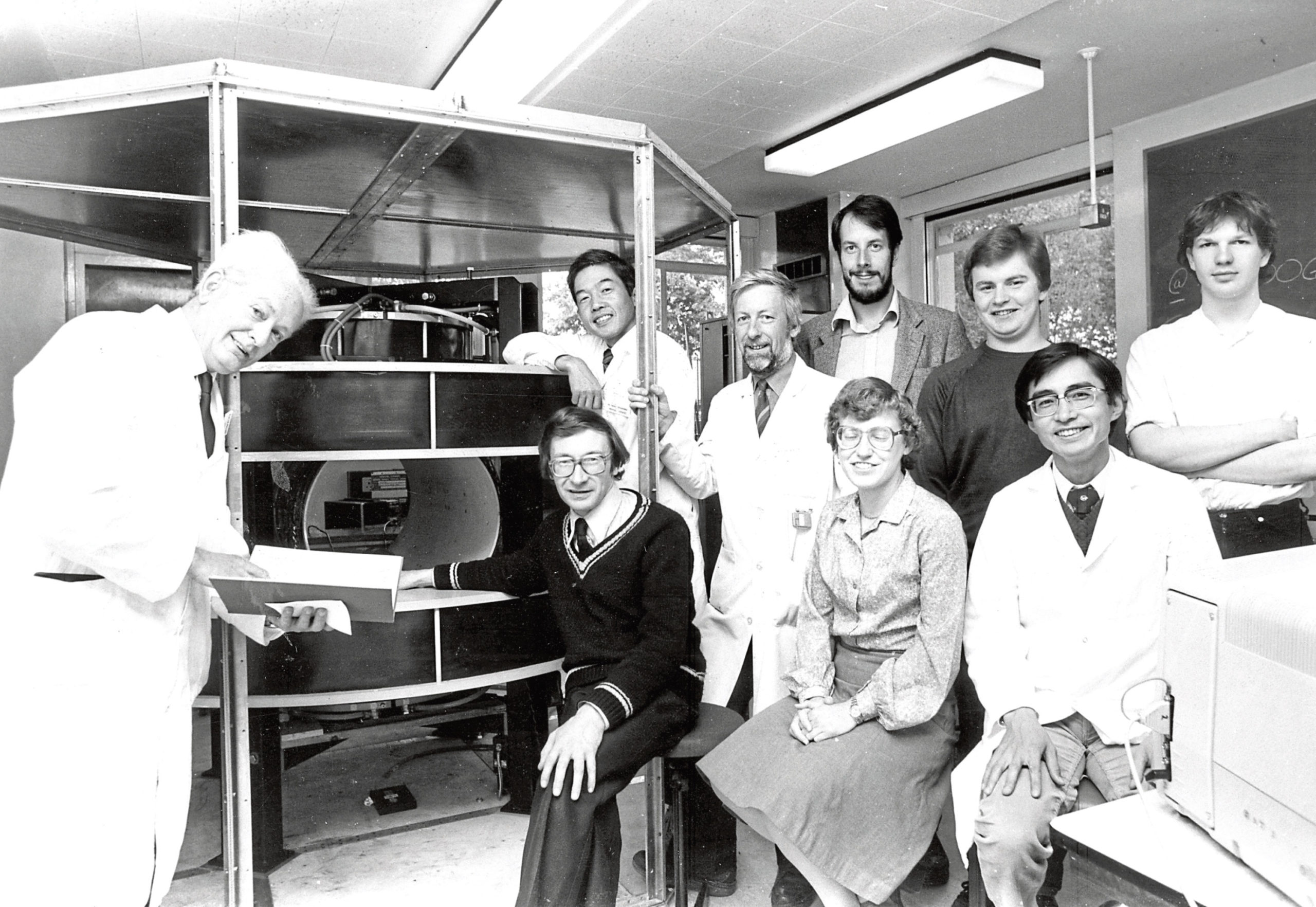
The machine was developed over 10 years by an Aberdeen University team, headed by Professor John Mallard and its inventor Dr James Hutchison.
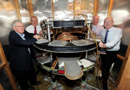
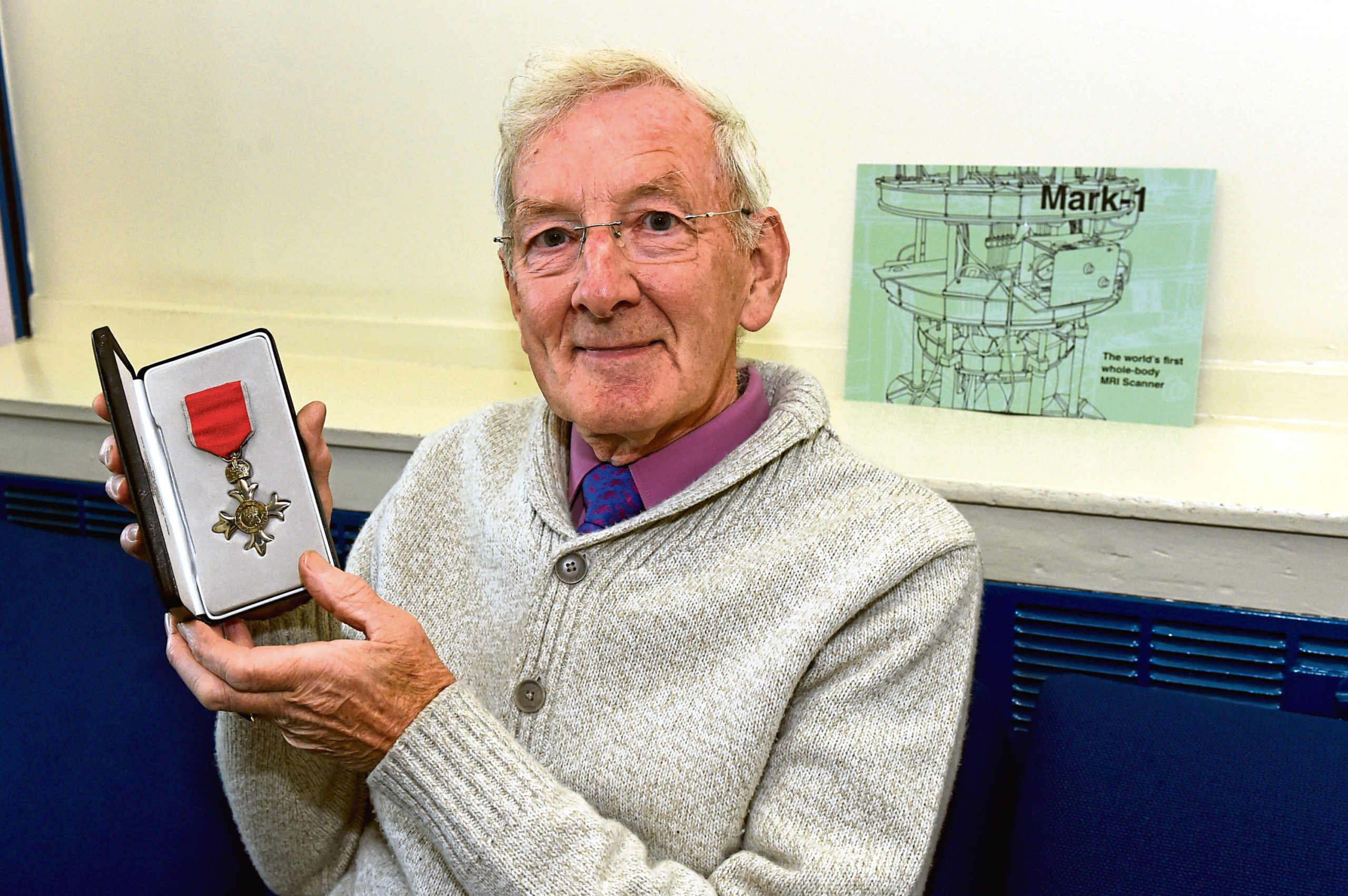
Edward “Eddie” Stevenson, 75, was one of the engineers in the medical physics department at the hospital who worked on bringing the machine to life.
He said: “At the time we didn’t know we were making history – it was just another day.”
As a senior clinical technologist, Eddie worked on adding water cooling to avoid overheating of patients on the hot magnet, building the tubing and creating the couch where the patient would be examined.
He added: “The technicians that actually built the MRI scanner Mark-1 and Mark-II were the late David Jack and Collin, our apprentice at the time. Without us, it would have never happened.”
Eddie, who spent 44 years working in the department, is also known for his Christmas display in his front garden on Ashgrove Road West and was awarded an MBE for his work with the Mark-1 and his charitable light display.
The machine was used to scan nearly 1,000 patients until it was replaced by a more powerful version, Mark-II, in 1983.
It was then used for research and teaching within Aberdeen University’s medical school before being put into storage in 2005.
The Mark-1 was rebuilt in 2016 and placed in the Suttie Arts Space inside Aberdeen Royal Infirmary for exhibition, where Eddie turned out to help the crew reassemble the pieces.
NHS Grampian’s head of imaging physics Dr Roger Staff said: “We were creating services here which were unique in the world. The evolution of MRIs were taken out of universities and big imaging companies started to invest into the field.
“And now imaging is almost an essential part of everything we do and it’s constantly growing.”
Meanwhile, a team from Aberdeen University has been leading the charge in recent years with the Fast Field Cycling (FFC) MRI Scanner.
MRI scanners use a large magnet along with pulses of radio waves to create detailed pictures of a patient’s anatomy.
However, the new FFC scanners are able to extract far more information by varying the strength of the magnetic field generated during the scanning procedure.
Group research leader Professor David Lurie said: “There’s been huge development in the hardware used for MRI and the software to produce the images.
“As for the hardware, one of the main things which has improved since the early days is strength of the magnetic field used which gives better signals for better quality images.
“Our project is at a similar stage to what was done with the original Mark-1 scanner in Aberdeen in the early 1980s.”
Around 25,000 MRI machines are now in daily use.
