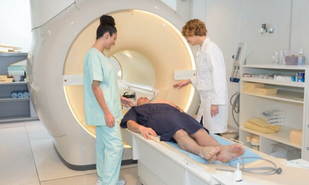
Using magnetic fields and radio waves to produce images of inside the body, the MRI scanner has been responsible for saving thousands of lives.
A medical breakthrough that took its first steps in the 1950’s, the scanner can examine almost any part of the human body and was the culmination of many leading scientists.
John Mallard led the team that created the first full-body MRI scanner here in Aberdeen.
Previous to his teams work, such equipment had only been used on mice and cadavers.
Now operated by radiographers in hospitals around the world, investigations into the human body have been made that much easier with his pioneering work into the technology.
Without involving X-rays or other radiation, images can be formed by MRI scanners that make use of radio waves and strong magnetic fields to generate images of inside the body.
The breakthroughs made by the Aberdeen University team in the 1970’s allowed for the first clinically useful image of a patient’s internal tissues using MRI.
Taking place in 1980 the machine identified a primary tumour in the patient’s chest, an abnormal liver, and secondary cancer in his bones.
These initial steps have allowed for results from an MRI scan to be used to help diagnose conditions, plan treatments and assess how effective previous treatment has been.
This machine was later used at St Bartholomew’s Hospital, in London, from 1983 to 1993.
Professor Mallard and his team are credited for the technological advances that led to the widespread introduction of MRI.
